
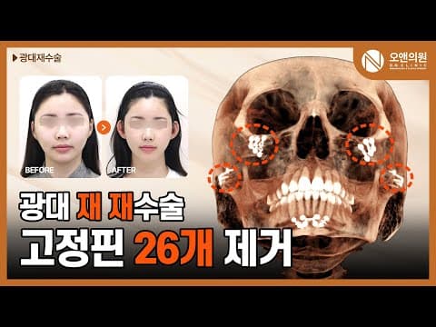
This patient, who visited On Clinic, had undergone zygoma surgery twice in the past but was dissatisfied with the results and presented with an astonishing 26 fixation pins in their face. The thinned zygoma and multiple surgical scars made revision surgery extremely challenging. However, On Clinic prioritized the patient's safety and aesthetic improvement, planning a delicate and precise customized zygoma revision surgery.
The Difficulty of Zygoma Revision Surgery and On Clinic's Approach
Analysis of the patient's 3D CT scans revealed that the zygoma had become abnormally thin due to previous surgeries, leading to a somewhat flat and broad facial appearance. Notably, the 26 pins used to fix the zygoma were conspicuously embedded throughout the face, and safely removing these pins alone required considerable time and precision.
Dr. Myungjun Oh thoroughly explained this complex situation during the consultation with the patient, emphasizing a safe surgical plan.
"We removed all the previously used pins and replaced them with a much smaller number of smaller-sized pins. Simply cutting doesn't cause severe bleeding, but resection does. If methods A and B yield the same result, one should choose the safer surgery. Why would anyone opt for a risky surgery? You should choose a safe surgery."
After removing the existing pins, bone grafting techniques were applied to reinforce the thinned zygoma and add three-dimensionality to the face. Utilizing the patient's own bone for grafting enhanced biocompatibility and aimed for a more natural result.
Securing Three-Dimensionality and Safety Through Bone Grafting
The goal was to improve the face, which had become broad due to the anterior zygoma being pushed backward by previous surgeries, by bringing it forward through bone grafting to create a convex and three-dimensional shape. Additionally, the right zygoma area, which was weakened by excessively thin bone, was reinforced with two fixation pins to enhance stability. Osteotomy of the coronoid process is performed to resolve issues with difficulty opening the mouth when the temporalis muscle is excessively developed. Through this complex approach, we aimed for functional and aesthetic improvements beyond simple reduction.
- Removal of Existing Pins: All 26 pins embedded in the face were removed to eliminate unnecessary foreign bodies.
- Bone Grafting: The thinned zygoma was reinforced by utilizing removed bone fragments or grafting the patient's own bone.
- Zygoma Repositioning: The anterior zygoma, which was pushed back, was moved forward to create a three-dimensional face shape.
- Secure Fixation: Fewer, smaller, and safer fixation pins were used compared to previous surgeries to reduce the risk of recurrence.
If post-surgical CT images are viewed only from the front, the bone shape might not appear significantly changed. However, On Clinic analyzes CT scans from various angles to capture even subtle changes. Particularly, a basal view CT image clearly shows that the zygoma, which was located outside the joint before surgery, narrowed inward towards the joint after surgery. These changes are visually noticeable, making the face appear smaller and more three-dimensional.
"When viewed from the front, you might mistakenly think the bone shape hasn't changed much. But from below, it's completely different. Now it's narrower than the joint. You can feel the reduction even by touching it!"
In many cases, when surgery is performed at other hospitals, too much bone is shaved, leaving the zygoma as thin as paper. Such weakened bone requires a more meticulous approach during revision surgery, and On Clinic focuses on reinforcing it through methods like bone grafting to enhance surgical safety and the longevity of the results.
Problems with Other Hospitals' Surgeries and On Clinic's Solutions
In this patient's case, because the zygoma was excessively thinned by previous surgeries, simply re-shaving the bone would not have yielded good results. Thin bones are difficult to fix, carry a high risk of recurrence, and can even fracture in severe cases. To address these issues, On Clinic performed bone grafting in necessary areas instead of removing unnecessary bone, thereby reinforcing the bone's thickness and strength. This approach allowed us to create a smooth and strong facial contour while achieving a more natural and three-dimensional face shape.
After the surgery, the patient expressed satisfaction, stating that their face had significantly reduced. On Clinic strives to meticulously analyze each patient's condition, even in such complex and challenging revision surgery cases, and provides optimal solutions based on the latest medical technology and extensive experience to achieve safe and satisfactory results.
Dr. Myungjun Oh, On Clinic | Last reviewed 2026-05-23